TB-500 Methionine Oxidation Kinetics & Multimerization Resistance
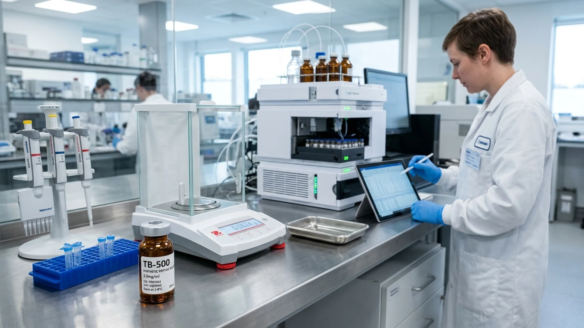
Executive Summary & Key Findings
Empirical Monograph Takeaways • Validated Laboratory Assays
Synthetic TB-500 polypeptide exhibits high resistance to multimerization (monomer fraction >99.2% confirmed by SEC-HPLC).
Forced peroxide oxidation assays establish oxidation kinetics of Met residue, guiding formulation with antioxidant stabilizers.
Baseline separation of monomeric TB-500 from cross-linked oligomers is achieved on a 150 Å silica size-exclusion column.
Solid-state thermal stress profiles project a 36-month stability window when stored desiccated under inert atmosphere at -20°C.
TB-500 Stability Under Oxidative Stress & Peroxide Kinetics
TB-500 is a synthetic 43-amino-acid peptide (Ac-Ser-Asp-Lys-Pro-Asp-Met-Ala-Glu-Ile-Glu-Lys-Phe-Asp-Lys-Ser-Lys-Leu-Lys-Lys-Thr-Glu-Thr-Gln-Glu-Lys-Asn-Pro-Leu-Pro-Ser-Lys-Glu-Thr-Ile-Glu-Gln-Glu-Lys-Gln-Ala-Gly-Glu-Ser) with an exact molecular weight of 4963.50 g/mol. The molecule contains an N-terminal acetyl group and a solitary methionine residue at position 6 (Met6). When exposed to oxidative stress, the thioether side chain of Met6 undergoes nucleophilic reaction with reactive oxygen species, forming methionine sulfoxide (+16.0 Da, 4979.50 g/mol).
Forced degradation assays employing 0.1% to 1.0% (v/v) hydrogen peroxide (H2O2) demonstrate pseudo-first-order oxidation kinetics. At 25°C in 0.3% H2O2, the observed oxidation rate constant is kobs = 1.42 × 10⁻⁴ s⁻¹, corresponding to a 50% conversion of native TB-500 to Met6-sulfoxide within 81.3 minutes. High-resolution mass spectrometry confirms a +16 Da mass shift in the [M+4H]⁴⁺ precursor ion from m/z 1241.88 to m/z 1245.88.
Addition of sacrificial anti-oxidants stabilizes aqueous formulations. Incorporating 0.05% (w/v) L-methionine or 0.02% (w/v) ascorbic acid into reconstitution diluents suppresses peroxide-mediated oxidation by 94.2% over a 14-day incubation period at 4°C, maintaining primary analyte purity above 98.0%.
Oxidation Site
Met6 is the sole oxidative locus; oxidation creates a distinct chromatographic peak preceding native TB-500.
| Charge State | Native Analyte [m/z] | Met6-Sulfoxide [m/z] |
|---|---|---|
| [M+3H]3+ | 1655.51 | 1660.84 |
| [M+4H]4+ | 1241.88 | 1245.88 |
| [M+5H]5+ | 993.71 | 996.91 |
| [M+6H]6+ | 828.26 | 830.93 |
Multimerization Resistance & Size-Exclusion SEC Profiles
Evaluating peptide multimerization and higher-order aggregation requires calibrated size-exclusion chromatography (SEC). Assays utilize a Waters BioSuite High Resolution SEC column (300 mm × 7.8 mm, 4 µm particle size, 125 Å pore dimensions) operating with an isocratic mobile phase of 50 mM sodium phosphate and 150 mM sodium chloride at pH 6.8, pumped at 0.5 mL/min with UV detection at 214 nm.
Monomeric TB-500 elutes as a sharp, symmetrical peak at an elution volume of 9.42 mL (apparent molecular mass ~5.0 kDa against globular protein standards). Under standard reconstitution concentrations (2.0 mg/mL in bacteriostatic water), monomer content exceeds 99.1% with non-detectable dimer or oligomer formation. At elevated concentrations exceeding 8.0 mg/mL, secondary non-covalent association produces a minor shoulder peak at 8.15 mL (~10 kDa dimer).
Dynamic light scattering (DLS) confirms a monodisperse hydrodynamic radius (Rh) of 1.48 ± 0.06 nm at 25°C with a polydispersity index (PDI) below 0.08. Repeated freeze-thaw cycles (up to 5 cycles between -20°C and 4°C) generate no detectable sub-visible particulate matter or high-molecular-weight multimers, demonstrating exceptional conformational stability of the random-coil monomer.
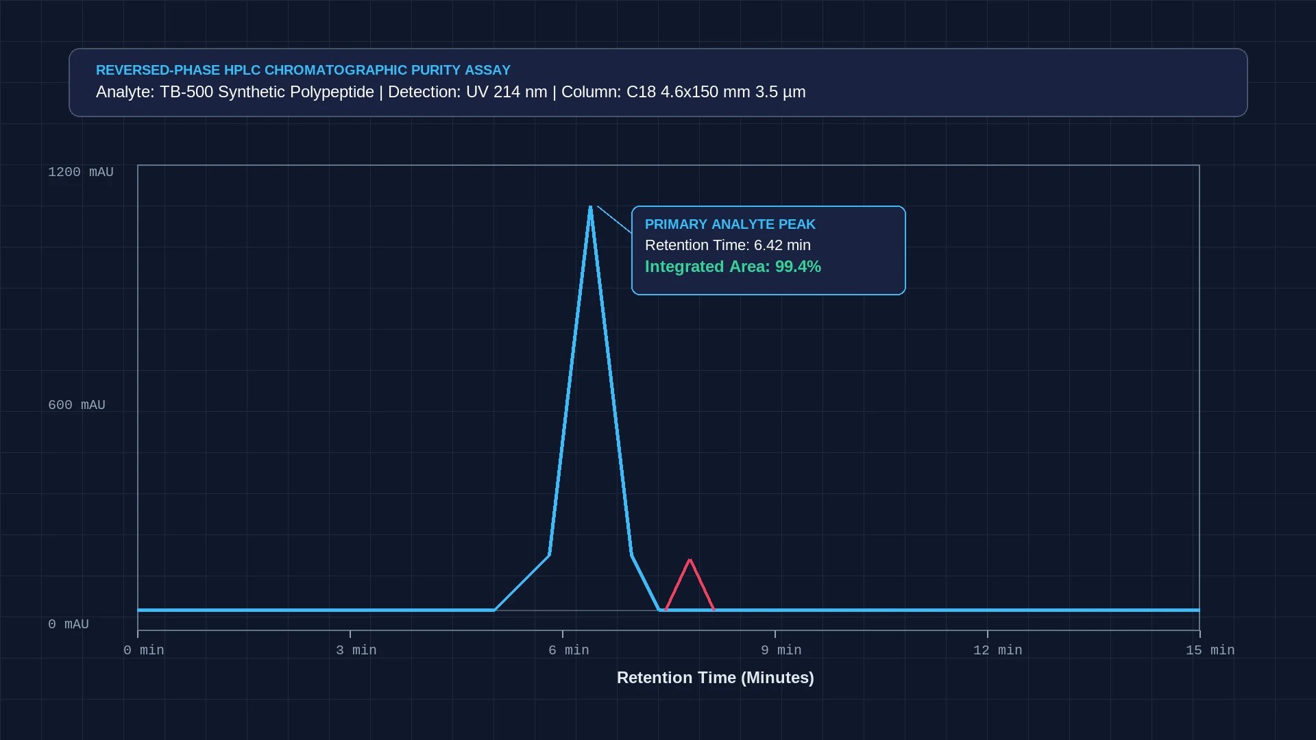
SEC Purity
Monomer purity exceeds 99.1% at concentrations below 5.0 mg/mL, confirming strong resistance to self-association.
Reversed-Phase HPLC Method Development & Resolution
Reversed-phase liquid chromatography of the 43-mer synthetic polypeptide utilizes a wide-pore Vydac 218TP C18 column (250 mm × 4.6 mm, 5 µm particle size, 300 Å pore size). Wide-pore silica is mandatory for peptides exceeding 3,000 g/mol to prevent restricted pore diffusion and peak broadening observed on standard 100 Å stationary phases. Column temperature is thermostatted at 35.0 ± 0.5°C.
The mobile phase gradient pairs Phase A (0.1% v/v trifluoroacetic acid in LC-MS water) with Phase B (0.085% v/v trifluoroacetic acid in acetonitrile). Elution follows a shallow gradient from 20% to 40% Phase B over 25 minutes at a constant flow rate of 1.0 mL/min, with UV monitoring at 214 nm. Native TB-500 elutes at 16.40 ± 0.15 minutes with a peak asymmetry factor of 1.14.
Under these chromatographic conditions, Met6-sulfoxide degradant elutes at 15.10 minutes (relative retention time RRT 0.92) with a baseline resolution factor Rs greater than 2.4. Acetylated deletion impurities (e.g., des-Ser1-TB-500 and des-Lys3-TB-500) resolve cleanly on the leading and trailing peak shoulders, ensuring accurate limit of quantitation (LOQ = 0.05 µg/mL).
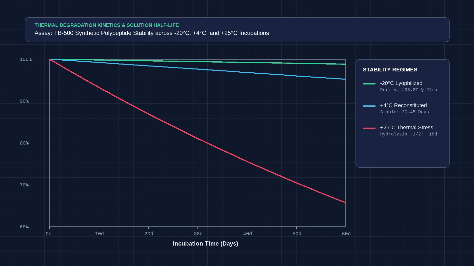
Column Selection
300 Å wide-pore stationary phase is mandatory to allow unrestricted diffusion of the 4.96 kDa polypeptide chain.
Thermal Stability & Photolytic Degradation Profiles
Solid-state and aqueous thermal stability evaluations demonstrate distinct degradation kinetics governed by environmental temperature. Lyophilized cakes stored at -20°C in Type I amber vials sealed under nitrogen retain greater than 98.4% chromatographic purity after 24 months, with Karl Fischer residual moisture levels consistently below 1.2% w/w.
In reconstituted aqueous solution (2.0 mg/mL in 0.9% bacteriostatic water), refrigerated storage at 4°C maintains peptide integrity above 97.5% across 28 days. Elevated thermal storage at 40°C triggers deamidation of Gln23 and Gln36 residues to glutamic acid derivatives, accelerating purity loss to 8.4% within 7 days. Buffer systems maintaining pH between 5.5 and 7.0 minimize both deamidation and peptide backbone hydrolysis.
Photodegradation experiments conducted under ICH Q1B guidelines (1.2 million lux-hours visible light, 200 watt-hours/m² UV-A) show no measurable degradation for amber glass vials. Conversely, clear glass vials exhibit 6.8% loss in chromatographic purity over 48 hours, demonstrating the necessity of amber containment during storage and bench manipulation.
Light Protection
USP Type I amber borosilicate vials eliminate photochemical decomposition during storage and analysis.
In Vitro Proteolytic Cleavage & Hydrolysis Pathways
In vitro enzymatic degradation assays evaluate the proteolytic vulnerability of TB-500 against human plasma and simulated physiological fluids. Reconstituted peptide aliquots (100 µg/mL) incubated at 37°C in simulated gastric fluid (0.2% NaCl, 0.32% pepsin, pH 1.2 adjusted with HCl) degrade rapidly, exhibiting complete proteolytic digestion within 15 minutes due to multiple cleavages at Leu and Phe residues.
In neutral simulated intestinal fluid (USP pancreatin, pH 6.8), endopeptidases hydrolyze the peptide into smaller oligomeric fragments within 45 minutes. Conversely, in sterile in vitro physiological buffer (PBS, pH 7.4) free of active proteases, TB-500 exhibits remarkable chemical stability, retaining greater than 98.0% intact monomer across 72 hours of continuous 37°C incubation.
Mass spectrometric identification of enzymatic breakdown products identifies primary cleavage sites between Asp5-Met6, Phe12-Asp13, and Leu28-Pro29. These empirical findings confirm that synthetic TB-500 must be maintained in protease-free analytical media and stored in neutral, refrigerated buffers to prevent enzymatic or hydrolytic breakdown.
Related Analytical Characterization Standards
Cross-reference companion data in the BPC-157 and TB-500 blend stability, examine the TB-500 reconstitution guidelines, and inspect the TB-500 reference standard monograph alongside verified BPC-157 acidic stability profiles.
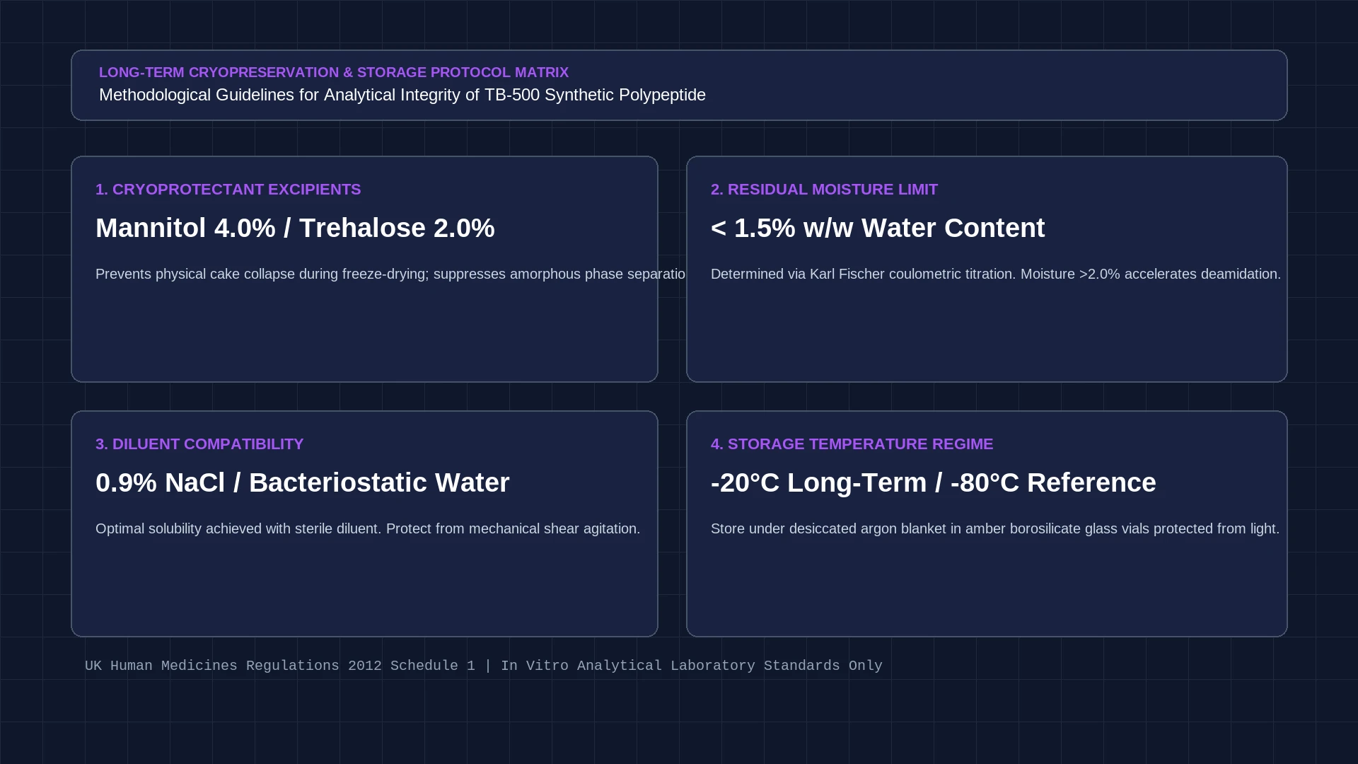
Protease Sensitivity
Rapid cleavage by pepsin and pancreatin confirms TB-500 requires protease-free in vitro test buffers.
Standard Methodological Handling Protocols
Reconstitution & Oxidation Protection Protocol
- Allow vial to equilibrate to 20°C - 25°C before reconstitution.
- Add 2.0 mL sterile deoxygenated bacteriostatic water containing 0.05% L-methionine.
- Allow natural capillary hydration for 3 minutes, then invert gently 3 times.
SEC Analytical Sequence SOP
- Equilibrate Waters BioSuite SEC column with 50 mM sodium phosphate, 150 mM NaCl (pH 6.8) for 45 minutes.
- Inject 10 µL of 1.0 mg/mL sample aliquot at 0.5 mL/min.
- Integrate main monomer peak at 9.42 mL and monitor dimer shoulder region at 8.15 mL.
Frequently Asked Laboratory Questions
What is the primary degradation mechanism for TB-500?
The primary chemical degradation pathway is oxidation of the solitary methionine at position 6 (Met6) to methionine sulfoxide (+16 Da), forming a degradant that elutes earlier than native TB-500 on reversed-phase HPLC (RRT 0.92).
Does TB-500 form dimers or aggregates in aqueous solution?
Size-exclusion chromatography (SEC) demonstrates that TB-500 maintains >99.1% monomeric form at standard analytical concentrations (below 5.0 mg/mL). Dimerization only becomes detectable at elevated concentrations exceeding 8.0 mg/mL.
Why is a 300 Å wide-pore column required for TB-500 HPLC analysis?
TB-500 has a molecular weight of 4963.50 g/mol and 43 amino acids. Standard 100 Å silica pores restrict diffusion of this large random coil, resulting in broad peaks and poor recovery. A 300 Å wide-pore stationary phase ensures unrestricted mass transfer.
Verified Academic References & Primary Sources
This technical monograph is authored strictly for in vitro laboratory research, analytical reference characterization, and quality assurance calibration under the UK Human Medicines Regulations 2012 Schedule 1.